What are Muscles?
Muscles are integral components of the human body, enabling movement, stability, and various essential bodily functions. Composed primarily of specialized cells called muscle fibers, muscles have unique properties that allow them to contract and generate force. This ability to contract is crucial for virtually every action we perform, from simple gestures like waving a hand to complex activities such as running or lifting weights. Muscles also play a significant role in maintaining posture and generating heat to keep our bodies warm. Given their vital functions, understanding the different types of muscles and how they operate is essential for anyone interested in fitness and health.
Types of Muscles
Skeletal Muscles
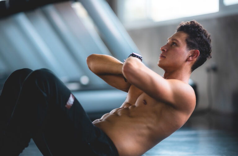
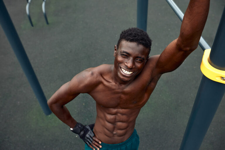
Skeletal muscles, also known as voluntary muscles, are attached to bones and are responsible for most of the movements we consciously control. These muscles are striated, meaning they have a banded appearance under a microscope, which results from their highly organized structure of muscle fibers. Each skeletal muscle fiber is a single cell containing multiple nuclei, allowing it to sustain prolonged and intense activity. These muscles work in pairs: while one muscle contracts, the other relaxes, enabling precise control of movement and posture. For example, when you bend your elbow, the biceps muscle contracts while the triceps muscle relaxes.
Skeletal muscles are adaptable and can increase in size and strength in response to regular exercise, particularly resistance training. This adaptability is known as muscle hypertrophy, a process driven by the stress and micro-damage inflicted on muscle fibers during exercise, which then repair and grow stronger. Additionally, skeletal muscles have a high metabolic rate, meaning they consume more energy at rest compared to other tissues, making muscle mass a critical factor in overall metabolic health and weight management.
Head and Neck Muscles
- Frontalis
- Temporalis
- Masseter
- Orbicularis oculi
- Orbicularis oris
- Buccinator
- Sternocleidomastoid
- Scalenes (Anterior, Middle, Posterior)
- Platysma
- Levator scapulae
- Trapezius (upper fibers)
Back Muscles
- Trapezius (middle and lower fibers)
- Latissimus dorsi
- Rhomboid major
- Rhomboid minor
- Levator scapulae
- Erector spinae (Iliocostalis, Longissimus, Spinalis)
- Multifidus
- Serratus posterior superior
- Serratus posterior inferior
- Quadratus lumborum
Chest Muscles
- Pectoralis major
- Pectoralis minor
- Serratus anterior
- Subclavius
- Abdominal Muscles
- Rectus abdominis
- External oblique
- Internal oblique
- Transversus abdominis
- Pyramidalis
Shoulder Muscles
- Deltoid (Anterior, Lateral, Posterior)
- Teres major
- Rotator Cuff Muscles
- Supraspinatus
- Infraspinatus
- Teres minor
- Subscapularis
Arm Muscles
- Upper Arm
- Biceps brachii
- Brachialis
- Triceps brachii
- Coracobrachialis
Forearm Muscles
- Brachioradialis
- Pronator teres
- Flexor carpi radialis
- Palmaris longus
- Flexor carpi ulnaris
- Flexor digitorum superficialis
- Flexor digitorum profundus
- Flexor pollicis longus
- Pronator quadratus
- Extensor carpi radialis longus
- Extensor carpi radialis brevis
- Extensor digitorum
- Extensor digiti minimi
- Extensor carpi ulnaris
- Supinator
- Abductor pollicis longus
- Extensor pollicis brevis
- Extensor pollicis longus
- Extensor indicis
Arm Muscles
- Upper Arm
- Biceps brachii
- Brachialis
- Triceps brachii
- Coracobrachialis
Forearm
- Brachioradialis
- Pronator teres
- Flexor carpi radialis
- Palmaris longus
- Flexor carpi ulnaris
- Flexor digitorum superficialis
- Flexor digitorum profundus
- Flexor pollicis longus
- Pronator quadratus
- Extensor carpi radialis longus
- Extensor carpi radialis brevis
- Extensor digitorum
- Extensor digiti minimi
- Extensor carpi ulnaris
- Supinator
- Abductor pollicis longus
- Extensor pollicis brevis
- Extensor pollicis longus
- Extensor indicis
Hand Muscles
Thenar Muscles
- Abductor pollicis brevis
- Flexor pollicis brevis
- Opponens pollicis
- Adductor pollicis
Hypothenar Muscles
- Abductor digiti minimi
- Flexor digiti minimi brevis
- Opponens digiti minimi
Leg Muscles
- Anterior Compartment
- Tibialis anterior
- Extensor digitorum longus
- Extensor hallucis longus
- Fibularis tertius
- Lateral Compartment
- Fibularis longus
- Fibularis brevis
- Posterior Compartment
- Gastrocnemius
- Soleus
- Plantaris
- Popliteus
- Flexor digitorum longus
- Flexor hallucis longus
- Tibialis posterior
Foot Muscles
- Dorsal Muscles
- Extensor digitorum brevis
- Extensor hallucis brevis
- Plantar Muscles
- Abductor hallucis
- Flexor digitorum brevis
- Abductor digiti minimi
- Quadratus plantae
- Lumbricals
- Flexor hallucis brevis
- Adductor hallucis
- Flexor digiti minimi brevis
- Plantar interossei
- Dorsal interossei
Smooth Muscles
Smooth muscles are found in the walls of internal organs such as the stomach, intestines, blood vessels, and the bladder. Unlike skeletal muscles, smooth muscles are not under voluntary control, meaning they operate automatically to perform essential bodily functions. These muscles are non-striated, which gives them a uniform, smooth appearance under a microscope. Smooth muscle cells are spindle-shaped and contain a single nucleus, and they contract more slowly than skeletal muscles but can sustain contractions for longer periods without fatigue.
The primary function of smooth muscles is to facilitate bodily processes such as digestion, blood flow, and the regulation of airway passages. For instance, smooth muscles in the walls of the intestines contract rhythmically to move food along the digestive tract, a process known as peristalsis. In blood vessels, smooth muscle contraction regulates blood pressure and flow by adjusting the diameter of the vessels. This type of muscle is highly resilient and can adjust its function in response to various physiological demands, ensuring the body’s internal environment remains stable.
Cardiac Muscle
The cardiac muscle is a specialized form of muscle found only in the heart. It shares some characteristics with both skeletal and smooth muscles. Like skeletal muscles, cardiac muscle is striated, which aids in its powerful contractions. However, similar to smooth muscles, cardiac muscle operates involuntarily, meaning it functions without conscious control to pump blood throughout the body continuously. This muscle type is composed of branching cells that connect tightly at intercalated discs, allowing rapid transmission of electrical impulses and synchronized contraction of the heart.
The rhythmic contractions of the cardiac muscle are regulated by the sinoatrial node, the heart’s natural pacemaker, which generates electrical signals that spread through the heart muscle. This coordination ensures the heart efficiently pumps blood, delivering oxygen and nutrients to tissues while removing waste products. The cardiac muscle is remarkably resilient and can function throughout a person’s lifetime without tiring, provided it remains healthy. Maintaining cardiovascular fitness through regular exercise, a balanced diet, and avoiding harmful habits like smoking is essential for supporting cardiac muscle health.
Anatomy of Muscles
Muscle Fiber Structure
Muscle fibers, the building blocks of muscles, are highly specialized cells designed for contraction and force generation. Each muscle fiber is encased in a plasma membrane called the sarcolemma, which helps transmit electrical signals initiated by nerve impulses. Inside the muscle fiber, the cytoplasm, known as sarcoplasm, contains numerous myofibrils – long, rod-like structures that run the length of the fiber. Myofibrils are composed of repeating units called sarcomeres, which are the functional units of muscle contraction.
Sarcomeres are made up of protein filaments called actin (thin filaments) and myosin (thick filaments). The interaction between these filaments, regulated by calcium ions and adenosine triphosphate (ATP), leads to muscle contraction. When a muscle fiber receives a signal from the nervous system, calcium ions are released within the sarcoplasm, initiating the binding of myosin to actin. This binding causes the filaments to slide past each other, shortening the sarcomere and generating contraction. This process, known as the sliding filament theory, is fundamental to all muscle contractions.
Muscle Groups and Attachments
Muscles are grouped based on their location and function within the body. Major muscle groups include the muscles of the upper body (such as the chest, back, and arms), the core muscles (including the abdominals and lower back), and the lower body muscles (such as the thighs, calves, and glutes). Each muscle group works in concert with others to produce coordinated movements and maintain stability. For instance, the muscles of the back and abdomen work together to support the spine and maintain posture.
Muscles are attached to bones by tendons, strong bands of connective tissue that transmit the force generated by muscle contractions to the skeleton. Tendons are highly resilient and can withstand significant stress, enabling powerful and precise movements. The points where tendons attach to bones are called origins and insertions. The origin is typically located on the bone that remains stationary during a movement, while the insertion is on the bone that moves. Understanding these attachment points is crucial for analyzing how muscles contribute to different movements and for designing effective training programs that target specific muscle groups.
Physiology of Muscle Contraction
Neuromuscular Junction and Signal Transmission
The neuromuscular junction is a critical site where the nervous system communicates with muscles to initiate contraction. It is the point where a motor neuron, a type of nerve cell, connects to a muscle fiber. The process begins when an electrical signal, or action potential, travels down the motor neuron to the neuromuscular junction. Upon arrival, the action potential triggers the release of a neurotransmitter called acetylcholine from the neuron’s terminal.
Acetylcholine diffuses across the synaptic cleft, the small gap between the neuron and muscle fiber, and binds to receptors on the muscle fiber’s sarcolemma. This binding generates an electrical signal within the muscle fiber, leading to the release of calcium ions from the sarcoplasmic reticulum, a specialized organelle within the muscle cell. The influx of calcium ions initiates the interaction between actin and myosin filaments, resulting in muscle contraction. This precise and rapid communication between the nervous system and muscles is essential for controlled and coordinated movements.
Mechanism of Contraction
The actual contraction of muscle fibers occurs through the sliding filament theory, which describes how actin and myosin filaments interact to shorten the sarcomere. When calcium ions are released within the muscle fiber, they bind to a protein called troponin, which is attached to the actin filaments. This binding causes a conformational change in another protein, tropomyosin, exposing binding sites on the actin filaments for the myosin heads to attach.
Myosin heads, powered by ATP, bind to the exposed sites on actin, forming cross-bridges. The myosin heads then pivot, pulling the actin filaments toward the center of the sarcomere. This movement, known as the power stroke, shortens the sarcomere and generates force. ATP is then used to detach the myosin heads from actin, allowing the cycle to repeat as long as calcium ions and ATP are available. This continuous cycle of cross-bridge formation, power stroke, and detachment underlies all muscle contractions, from a gentle touch to a powerful lift.
Types of Contractions
Muscles can contract in different ways depending on the type of movement and the load being handled. Isometric contractions occur when muscle tension is generated without a change in muscle length. This type of contraction is common in activities that require maintaining a static position, such as holding a plank or carrying a heavy object without moving. Isometric exercises are effective for building strength and endurance without joint movement.
Isotonic contractions, on the other hand, involve a change in muscle length while maintaining constant tension. There are two types of isotonic contractions: concentric and eccentric. Concentric contractions occur when the muscle shortens as it generates force, such as when lifting a weight during a bicep curl. Eccentric contractions happen when the muscle lengthens under tension, such as when lowering the weight back down. Both types of isotonic contractions are important for overall muscle development and functional strength.
Muscle Growth and Repair
Muscle Hypertrophy
Muscle hypertrophy refers to the increase in muscle size resulting from regular resistance training or other forms of exercise that place stress on the muscles. This process begins with the mechanical overload of muscle fibers during exercise, causing micro-damage to the tissue. In response, the body initiates a repair process that not only heals the damaged fibers but also adds new protein filaments to strengthen them. This adaptive response is driven by several factors, including the release of growth factors, hormones like testosterone and growth hormone, and increased protein synthesis.
Effective muscle hypertrophy requires a combination of progressive overload, where the intensity of the exercise is gradually increased, and adequate nutrition, particularly sufficient protein intake. Consistency in training and proper recovery periods are also crucial. During recovery, muscle fibers repair and grow stronger, preparing them for future challenges. Understanding the principles of muscle hypertrophy helps in designing training programs that maximize muscle growth and strength.
Muscle Recovery and Repair
The recovery and repair process is vital for muscle growth and overall performance. After intense exercise, muscles need time to repair the micro-damage sustained during training. This repair process involves satellite cells, a type of stem cell located on the surface of muscle fibers. When activated by muscle damage, satellite cells proliferate and fuse with the existing muscle fibers, contributing to their growth and repair.
Adequate rest, nutrition, and hydration are essential components of muscle recovery. Protein plays a crucial role in repairing muscle fibers, while carbohydrates replenish glycogen stores depleted during exercise. Additionally, rest allows the body to carry out necessary repair processes and reduces the risk of overtraining and injury. Incorporating rest days and varying the intensity of workouts can help maintain a balance between training and recovery, optimizing muscle health and performance.
Impact of Aging on Muscle Mass
As we age, muscle mass and strength tend to decline, a condition known as sarcopenia. This natural process can begin as early as the third decade of life and accelerates with age. Factors contributing to sarcopenia include hormonal changes, reduced physical activity, and diminished protein synthesis. The loss of muscle mass can lead to decreased strength, mobility issues, and an increased risk of falls and fractures.
However, regular resistance training and physical activity can significantly mitigate the effects of aging on muscle mass. Strength training stimulates muscle protein synthesis and helps maintain muscle size and function. Additionally, adequate protein intake and proper nutrition support muscle health. By staying active and incorporating strength training into their routines, older adults can maintain muscle mass, improve quality of life, and reduce the risk of age-related muscle decline.
Muscular Health and Diseases
Preventing Muscular Diseases
Maintaining muscular health involves a combination of regular physical activity, balanced nutrition, and lifestyle choices that support overall well-being. Regular exercise, particularly strength training and aerobic activities, helps preserve muscle mass and function. Additionally, a diet rich in protein, vitamins, and minerals supports muscle repair and growth. Hydration is also critical, as muscles require adequate fluid balance to function optimally.
Avoiding harmful habits such as smoking and excessive alcohol consumption can also protect muscle health. Smoking has been linked to reduced muscle strength and mass, while excessive alcohol intake can impair protein synthesis and muscle recovery. Ensuring adequate sleep is another important factor, as restorative sleep allows the body to carry out essential repair processes.
Common Muscular Disorders
Muscular disorders can affect the function and health of muscles, leading to various symptoms and complications. Some common muscular disorders include muscular dystrophy, myasthenia gravis, and inflammatory myopathies. Muscular dystrophy encompasses a group of genetic disorders characterized by progressive muscle weakness and degeneration. Myasthenia gravis is an autoimmune disorder that impairs communication between nerves and muscles, leading to muscle weakness and fatigue. Inflammatory myopathies, such as polymyositis and dermatomyositis, involve chronic inflammation of the muscles, resulting in weakness and damage.
These conditions often require medical intervention and specialized care to manage symptoms and maintain muscle function. Physical therapy, medications, and in some cases, surgery, can help improve the quality of life for individuals with muscular disorders. Early diagnosis and appropriate treatment are crucial for managing these conditions effectively.
Role of Exercise in Muscular Health
Exercise plays a pivotal role in maintaining and improving muscular health. Regular physical activity stimulates muscle growth, enhances strength, and improves endurance. Strength training, in particular, is effective for increasing muscle mass and strength. Exercises such as weightlifting, resistance band exercises, and bodyweight exercises target specific muscle groups and promote muscle hypertrophy.
Endurance training, such as running, cycling, and swimming, improves cardiovascular fitness and enhances the efficiency of muscle function. Additionally, flexibility exercises, including stretching and yoga, help maintain the range of motion and prevent injuries. A well-rounded exercise routine that incorporates strength, endurance, and flexibility training supports overall muscular health and reduces the risk of chronic diseases.
Muscle Fitness and Training
Strength Training
Strength training is a fundamental component of fitness that focuses on increasing muscle strength and size through resistance exercises. These exercises involve working against a force, such as free weights, resistance bands, or body weight. Strength training exercises include squats, deadlifts, bench presses, and rows, targeting major muscle groups. Proper technique and form are essential to prevent injuries and maximize the benefits of strength training.
Progressive overload is a key principle in strength training, involving gradually increasing the weight, resistance, or intensity of exercises to continuously challenge the muscles. This progression stimulates muscle adaptation, leading to increased strength and hypertrophy. Incorporating a variety of exercises and varying the intensity can help prevent plateaus and promote balanced muscle development.
Endurance Training
Endurance training focuses on improving the ability of muscles to sustain prolonged activity and resist fatigue. This type of training typically involves aerobic exercises such as running, cycling, swimming, and rowing. These activities increase the heart rate and enhance cardiovascular fitness, allowing muscles to work more efficiently for extended periods.
Endurance training also promotes the development of slow-twitch muscle fibers, which are more resistant to fatigue and capable of sustained contractions. Incorporating interval training, which alternates between high-intensity bursts and lower-intensity recovery periods, can further enhance endurance and cardiovascular health. Consistency and gradual progression are important for improving endurance and achieving long-term fitness goals.
Flexibility and Muscle Balance
Flexibility is an important aspect of overall fitness that involves the ability of muscles and joints to move through their full range of motion. Regular stretching exercises help maintain flexibility, reduce muscle stiffness, and prevent injuries. Activities such as yoga and Pilates are effective for improving flexibility and promoting muscle balance.
Maintaining muscle balance involves ensuring that opposing muscle groups are equally strong and flexible. Imbalances can lead to poor posture, decreased performance, and a higher risk of injuries. Incorporating exercises that target both the agonist (primary mover) and antagonist (opposing) muscles helps maintain muscle balance and overall functional fitness. Regular assessments and adjustments to the training program can help address any imbalances and support optimal muscle function.
Key Takeaways
Muscles are essential components of the human body, enabling movement, stability, and various bodily functions. Understanding the different types of muscles, their anatomy, and physiology is crucial for maintaining muscular health. Regular exercise, proper nutrition, and healthy lifestyle choices support muscle growth, repair, and overall function. Addressing muscular health and fitness can lead to improved quality of life, reduced risk of chronic diseases, and enhanced physical performance.